There is a particular kind of frustration that patients with breast tuberculosis carry by the time they finally reach a specialist. Most of them have already been told, at various points, that their lump is an abscess, then an infected cyst, then possibly cancer. They have had antibiotics that did not work. They have had incisions that did not heal. Some have been waiting for a biopsy result that came back inconclusive because the right tests were never ordered.
Breast tuberculosis sits in a diagnostic blind spot. It is uncommon enough that it is not the first thing a clinician considers, but common enough in the Indian subcontinent that missing it carries real consequences. Dr. Rohan Khandelwal, who practices across Delhi NCR and Gurgaon, sees this pattern repeatedly, and the defining feature is not the condition itself, but how long it went unrecognised.
The Biology Behind Why Breast TB Behaves the Way It Does
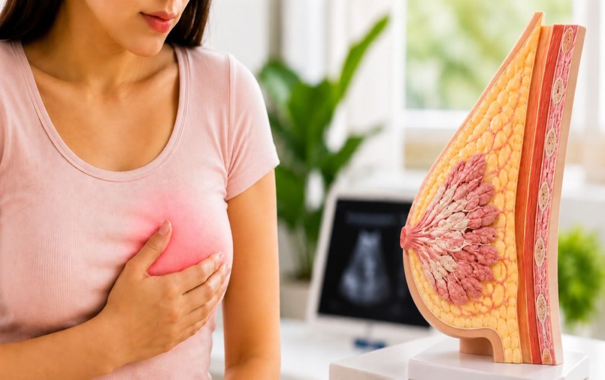
Breast tuberculosis seldom begins in the breast. Mycobacterium tuberculosis reaches breast tissue via the lymphatics or bloodstream, typically from a primary focus elsewhere in the body, such as the lungs, lymph nodes, or spine. In the majority of patients, the primary focus has already healed or is entirely subclinical. There is no cough, no X-ray finding, no history of TB that a patient would think to mention.
Once established in breast tissue, the infection creates a granulomatous reaction. This is the body's attempt to wall off an organism it cannot easily destroy. The result is a hard, irregular lump that is indistinguishable on feel from a malignancy. Over time, the centre of the granuloma may soften and liquefy, producing a cold abscess that can eventually track to the skin surface and open as a discharging sinus. At this stage, the wound will not close with standard wound care because the underlying infection is still active.
Breast tuberculosis causes are more prevalent in India is not just the higher background TB burden but also the fact that malnutrition, overcrowding, and immunological factors create a larger pool of people with latent or subclinical TB who may reactivate under physiological stress. Pregnancy, breastfeeding, and hormonal fluctuations in the breast are all factors that may play a role in localising reactivation to breast tissue.
Three Presentations That Are Routinely Misread
The clinical picture of breast TB does not follow a single script. Broadly speaking, there are three dominant presentations, and each tends to be misdiagnosed as something different.
- The hard, irregular lump without skin changes. This is the presentation most frequently mistaken for breast cancer. It is firm, often fixed to surrounding tissue, and does not feel like a benign fibroadenoma. Without a high index of suspicion for TB, a biopsy may be sent for routine histopathology without requesting the specific stains or cultures needed to identify mycobacteria.
- The fluctuant swelling with skin redness. This presentation is almost universally treated as a pyogenic abscess. Antibiotics are prescribed; when they fail, incision and drainage is performed. The wound does not heal. The patient is referred further along, still without a breast TB diagnosis.
- The chronic discharging sinus. By this stage, the disease has been present for months. The sinus tract from the original abscess has opened to the skin and continues to discharge thin, non-purulent fluid. This pattern is pathognomonic for TB if the clinician is thinking about it, but it is frequently managed as a surgical wound problem without the underlying cause being addressed.
Why Standard Tests Miss It
A routine culture of breast abscess pus will grow common bacteria if there is co-infection, or grow nothing if the TB bacilli are present in low numbers. Standard histopathology may report granulomas, but label them as non-specific without AFB staining being requested. The Mantoux test has limitations in a country where BCG vaccination is universal, producing false positives. And chest X-rays are often normal, which falsely reassures both patient and clinician that TB is not the diagnosis.
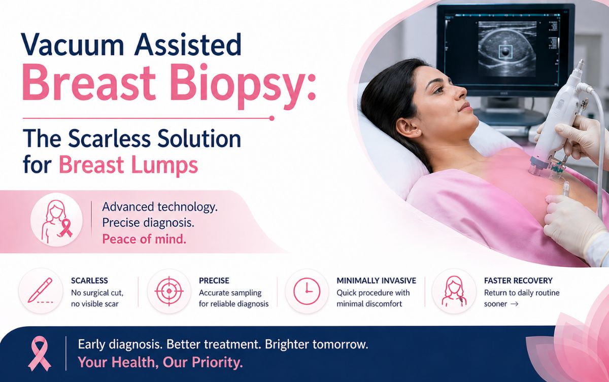
The modern diagnostic approach is considerably more reliable. Ultrasound-guided core needle biopsy collects an adequate tissue sample with minimal trauma. This sample is then sent simultaneously for histopathology with Ziehl-Neelsen staining, mycobacterial culture on Lowenstein-Jensen medium, and CBNAAT (GeneXpert), which can detect TB DNA and rifampicin resistance within a few hours. This combination gives a high diagnostic yield and eliminates the month-long wait for culture results that previously delayed treatment.
| Test | What It Detects | Limitation |
| AFB smear | Mycobacteria in tissue | Low sensitivity in small samples |
| Mycobacterial culture | Live organisms, drug sensitivity | Results take 4 to 8 weeks |
| CBNAAT / GeneXpert | TB DNA + rifampicin resistance | Does not test all drug resistances |
| Histopathology with ZN stain | Granulomas, caseous necrosis | Granulomas can have other causes |
| GRA (Interferon Gamma Release) | Immune response to TB antigens | Cannot distinguish active from latent TB |
The Actual Cost of a Late Diagnosis
A patient who receives the correct diagnosis in the first two months faces six months of oral anti-tubercular therapy and full recovery with no surgical intervention in most cases. A patient who is diagnosed after six months of mismanagement may have a large abscess cavity, significant skin involvement, one or more sinus tracts, and potential scarring of breast tissue that is difficult to reverse.
Beyond the physical toll, there is the financial cost of repeated consultations, failed antibiotic courses, ultrasound-guided drainage procedures, and wound management. There is also the psychological cost; the persistent anxiety of a patient who has been told different things by different doctors and still has no clear answer.
Anti-tubercular therapy for breast TB follows the same regimen used for pulmonary TB: an intensive phase of four drugs for two months, followed by a continuation phase of two drugs for four to seven months. Response is monitored clinically and radiologically. In most patients, the lump begins to shrink within six to eight weeks and resolves completely by the end of treatment.
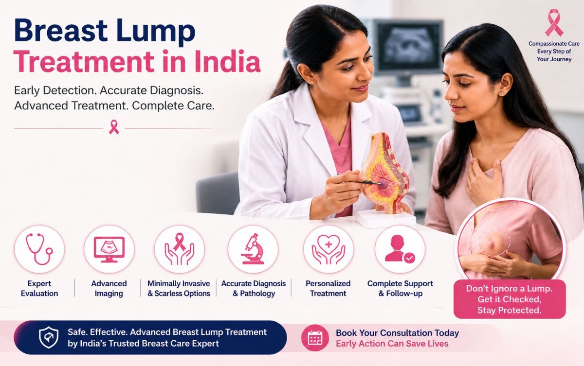
Getting the Right Evaluation
If you have a breast lump that has not been satisfactorily explained, a wound that keeps recurring, or a history of TB contact with a new breast symptom, the evaluation needs to go beyond a routine ultrasound and antibiotic course. Dr. Rohan Khandelwal sees patients across Gurgaon and the Delhi NCR region and has experience managing complex breast conditions where the initial diagnosis was incomplete or incorrect.
Frequently Asked Questions
No. Breast tuberculosis is localised to breast tissue and is not transmitted through physical contact, shared living spaces, or breastfeeding. It does not carry the same transmission risk as pulmonary TB.
Recurrence is uncommon if the full course of anti-tubercular therapy is completed. Incomplete treatment is the primary risk factor for relapse and for the development of drug-resistant TB.
No. A significant proportion of patients with extrapulmonary TB, including breast TB, have no active lung disease and a completely normal chest X-ray. The diagnosis is made through breast tissue biopsy and targeted microbiological testing.
Surgery is reserved for specific situations: a large abscess requiring drainage, a persistent sinus tract that has not closed after completing ATT, or a residual mass requiring excision. It is never the first intervention and should always follow a confirmed microbiological diagnosis.