Why Fibroadenomas Are So Common in Young Indian Women
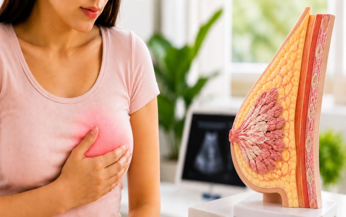
The average age at which a fibroadenoma is first detected in Indian women is significantly younger than the global average. Many patients are in their late teens or early to mid-twenties when they notice a smooth, movable lump that sends them into a spiral of anxiety, internet searches, and an urgent need for answers that often leads to conflicting information.
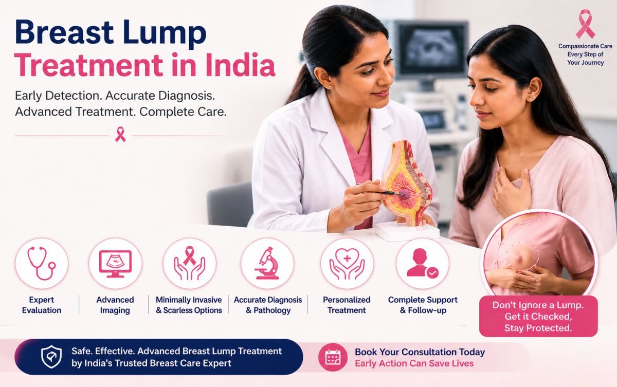
The standard clinical advice, confirm it is benign, monitor it, and leave it alone unless it grows, is medically sound for many cases. What it does not always address is the lived experience of carrying a lump you were told is harmless, but which you think about every day. Dr. Rohan Khandelwal, the best doctor for fibroadenoma in Delhi who specialises in breast conditions across Gurgaon and Delhi NCR, sees this reality in his practice constantly. The conversation around fibroadenoma has moved well past the basic question of whether it is cancerous. The more relevant questions are different.
Why Fibroadenomas Are So Common in Young Indian Women
Fibroadenomas are oestrogen-sensitive structures. They form, grow, and sometimes shrink in direct response to hormonal fluctuations. This is why they most commonly appear during the reproductive years, tend to enlarge during pregnancy, and often regress spontaneously after menopause.
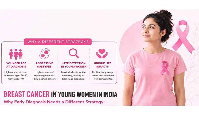
In India, the relatively early onset of regular menstrual cycles, combined with widespread nutritional variation and a high prevalence of hormonal irregularities, creates a fertile environment for fibroadenoma formation. It is also worth noting that many Indian women notice lumps only when they are already moderately sized, partly because routine breast self-examination is not yet part of standard health education for young women in many communities.
Multiple fibroadenomas, where a patient has two or more lumps either in the same breast or bilaterally, are not unusual. The presence of multiple fibroadenomas does not increase malignancy risk, but it does complicate management decisions and makes specialist review even more important. Consult Dr. Rohan Khandelwal for scarless fibroadenoma surgery.
The Lump That Kept Growing — When Monitoring Becomes the Wrong Choice
A significant subset of fibroadenomas does not stay stable. They grow. In some patients, a lump that was 1 cm at first detection becomes 2.5 cm within a year. This pattern, sometimes described as a juvenile or cellular fibroadenoma, is more common in adolescents and young adults and tends to have a more proliferative biology than the typical adult-type fibroadenoma.
The conventional threshold for intervention is typically a lump larger than 3 cm, or one that has shown documented growth over successive imaging studies. But this threshold is not absolute. A patient with a 2 cm fibroadenoma that has grown rapidly in six months may warrant removal sooner than a patient with a stable 2.8 cm lump. These decisions require clinical judgement, not just a size cutoff.
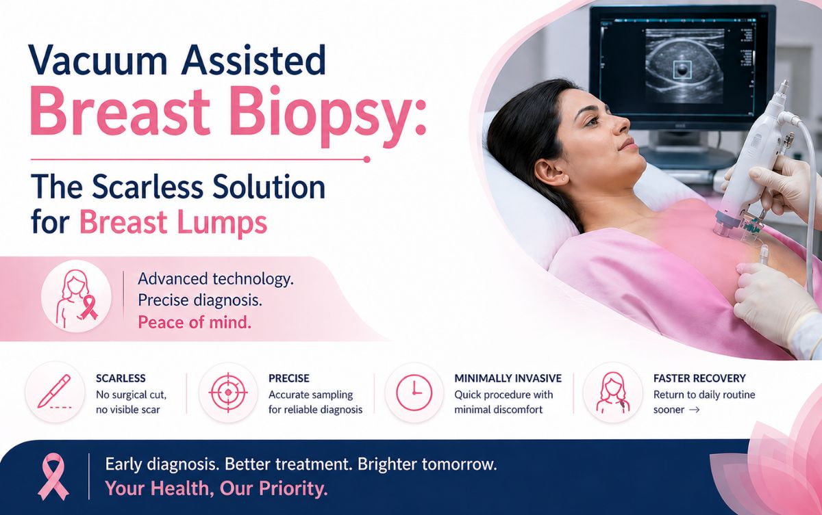
What has changed in recent years is that the availability of vacuum assisted excision has shifted the calculus. When removal required open surgery with its attendant scar, recovery, and surgical risk, watchful waiting was often the more proportionate choice for borderline cases. When removal can be achieved through a 3 mm entry point under local anaesthesia with a same-day return home, the calculus changes meaningfully for both patient and clinician.
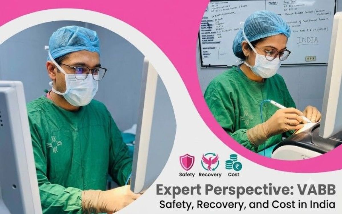
What the Vacuum Assisted Procedure Actually Involves — Beyond the Summary
Vacuum-assisted breast biopsy (VABB) uses a handheld probe connected to a vacuum system, operated under continuous real-time ultrasound imaging. The probe is inserted through a small nick in the skin, positioned adjacent to the fibroadenoma, and then activated. The vacuum draws the lump tissue into the cutting aperture in sequential passes, removing it in cores that are collected, labelled, and sent entirely for histopathology.
The precision of ultrasound guidance means the procedure is targeted directly at the lump with minimal disturbance to surrounding glandular tissue. There is no cavity left behind that needs packing or suturing. The small entry point is closed with a steri-strip and covered with a dressing. The patient leaves the procedure room the same day, typically within an hour of arriving.
| Parameter | VABE Procedure | Open Surgical Excision |
|---|---|---|
| Setting | Outpatient, procedure room | Operation theatre |
| Anaesthesia | Local injection only | General or spinal |
| Skin entry | 3 to 4 mm nick | 2 to 4 cm incision |
| Scar outcome | Imperceptible after healing | Visible scar |
| Return to work | The next day, for most patients | 5 to 10 days |
| Histopathology | Full tissue analysis done | Full tissue analysis done |
| Multiple lumps in one session | Often possible | Each requires a separate incision |
The Question of Multiple Fibroadenomas
One of the genuinely underappreciated advantages of vacuum assisted excision over open surgery is the ability to address multiple fibroadenomas in a single sitting. A patient with three small lumps in the same breast, or lumps in both breasts, faces a very different surgical equation with open surgery, multiple incisions, multiple scars, a longer procedure, and greater tissue disruption. With vacuum assisted excision fibroadenoma, multiple lumps can often be approached through the same or adjacent entry points during a single session, reducing both procedure time and total trauma to the breast.
This is particularly relevant for the subset of Indian patients who present with bilateral or multiple fibroadenomas and who may have deferred seeking treatment precisely because they believed removal would involve significant surgery.
What Supportive Therapy Can and Cannot Do
There is interest, particularly in India, in conservative or supportive management of fibroadenoma. Dietary changes, hormonal supplements, and certain anti-oestrogen preparations have been studied with variable results. The honest clinical answer is that supportive therapy can reduce associated breast discomfort and may slow the rate of growth in some cases, but it does not reliably shrink or eliminate an established fibroadenoma. Patients who have been told their lump will dissolve with medication should ask for imaging evidence of that claim at follow-up. Consult fibroadenoma specialist Dr. Rohan Khandelwal for consultation.
Having the Right Conversation
A fibroadenoma consultation is not just about deciding whether to remove a lump. It is a conversation about the patient's age, plans, anxiety level, lump behaviour over time, and which option carries the best balance of benefit and burden for that individual. For patients across Gurgaon, Delhi NCR, and Haryana, Dr. Rohan Khandelwal offers this conversation based on current evidence and extensive procedural experience.
Frequently Asked Questions
The same hormonal environment that produced one fibroadenoma can produce another, in the same breast or the opposite one. Having had a fibroadenoma is a risk factor for future fibroadenomas, though not for malignancy.
Intervention during pregnancy is generally deferred unless the lump is growing rapidly or causing significant discomfort. Many fibroadenomas enlarge during pregnancy and regress after delivery. Dr. Rohan Khandelwal advises review based on each patient's specific situation.
There is no absolute upper size limit, but fibroadenomas above 3 cm are generally considered candidates for removal. Giant fibroadenomas, defined as those above 5 cm, almost always require excision regardless of symptoms.
No. The breast tissue naturally fills in after the lump is removed. Any initial subtle contour change resolves within weeks as the surrounding tissue reorganises.